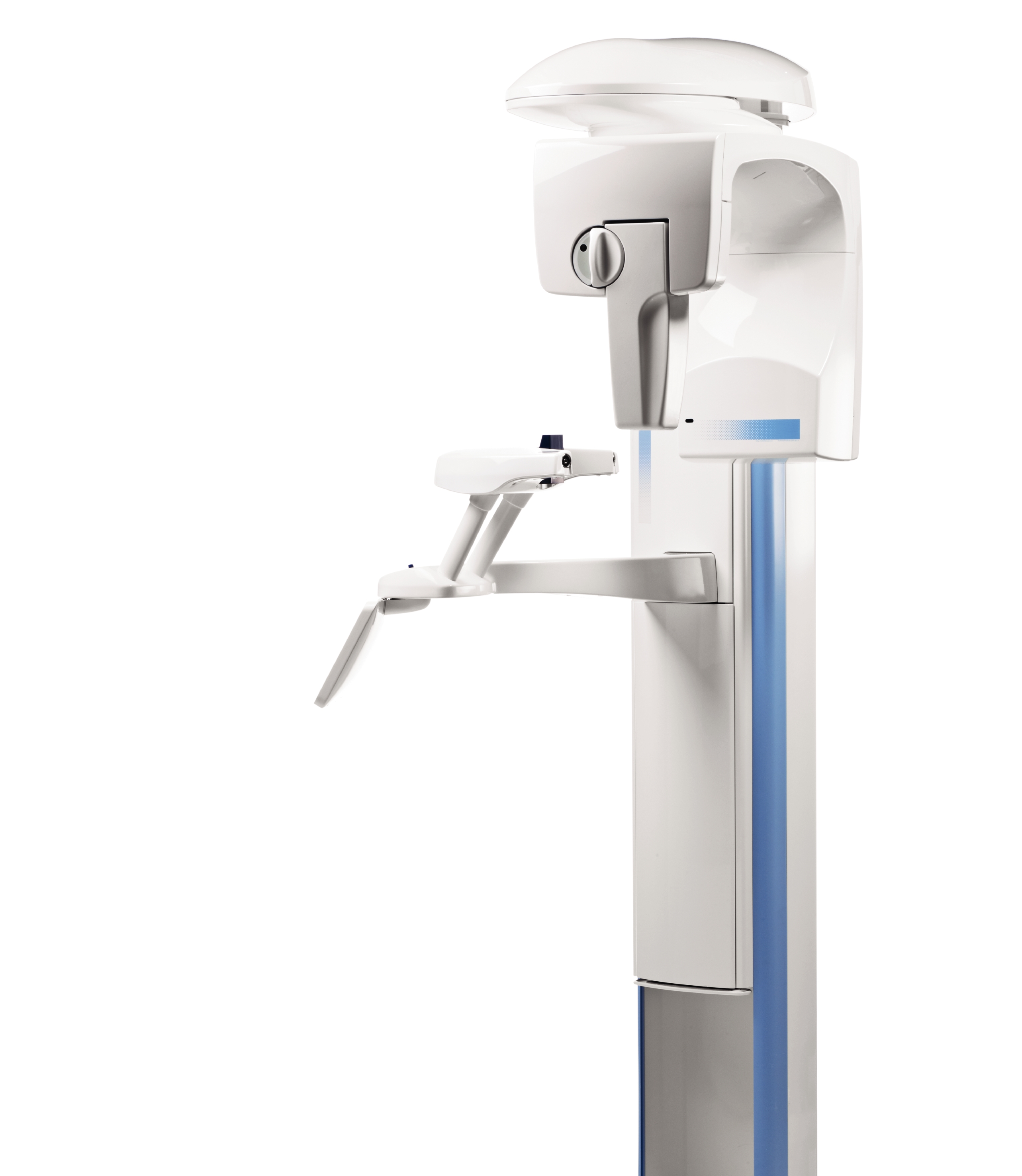
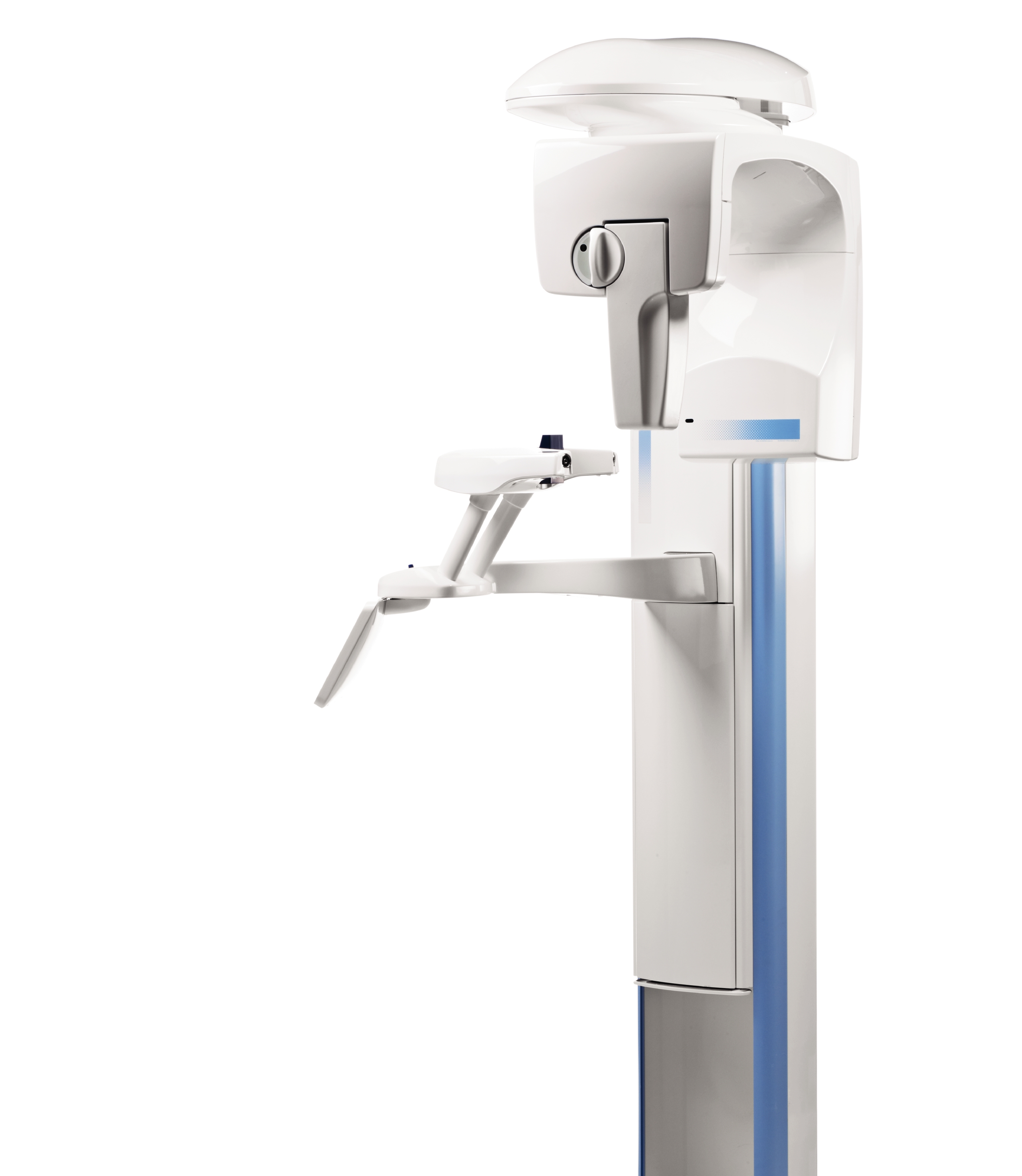
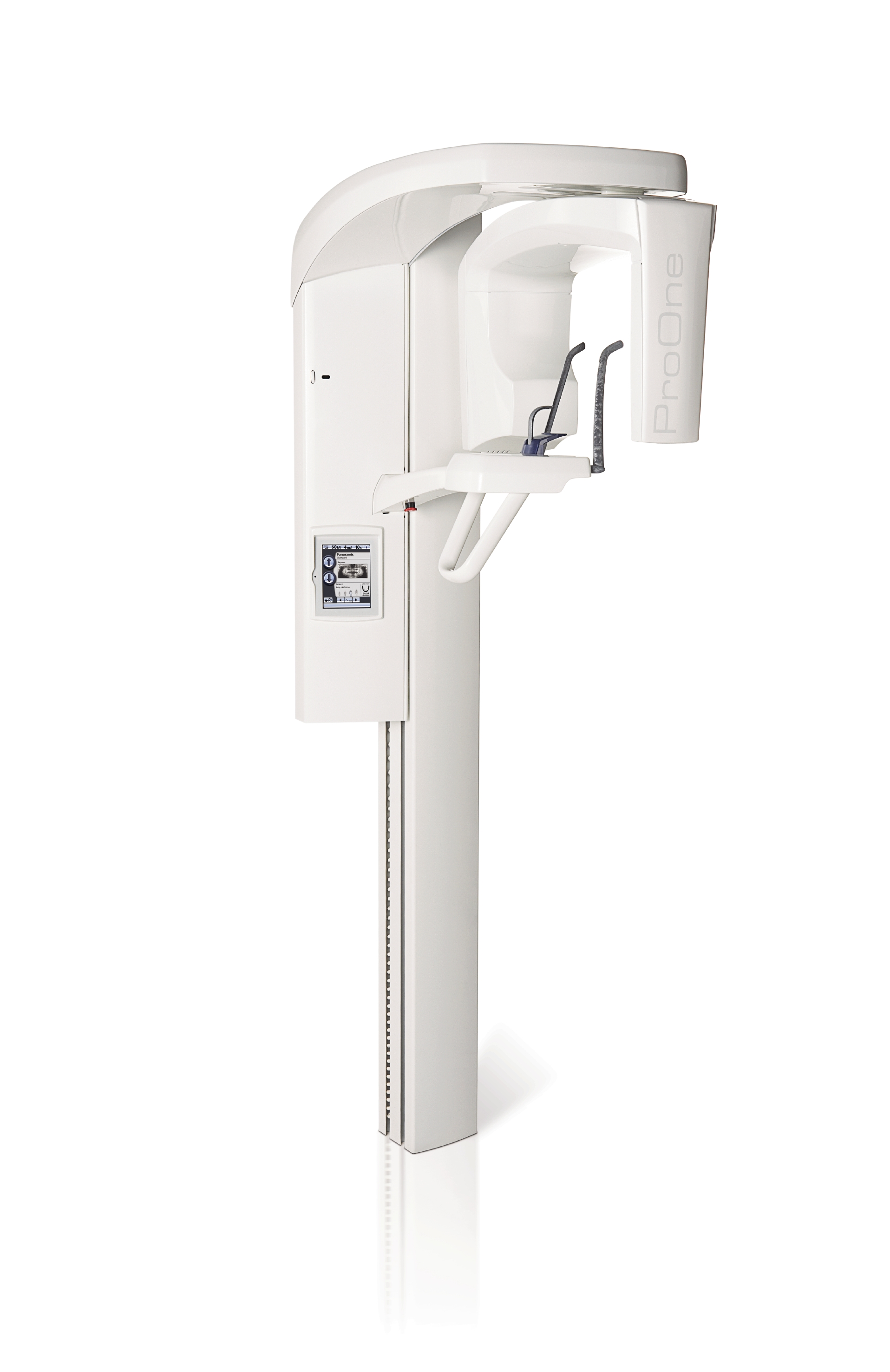
A complete maxillofacial imaging system.
Easy patient positioning
Open patient positioning and side entry minimises errors caused by incorrect patient positioning by allowing you to monitor the patient freely from both the front and side. Side entry allows easy access for all patients – standing or seated. Patient positioning is assisted by the triple laser beam system, which indicates the correct anatomical positioning points.
User interface provides guidance
The full-colour graphical user interface provides clear texts and symbols to guide you through your procedure. Settings are logically grouped and easy to understand, speeding up imaging and allowing you to focus on positioning your patient correctly and communicating with them.
Autofocus- for perfect panoramics every time
The unique autofocus feature automatically positions the focal layer using a low-dose scout image of the patient’s central incisors. Landmarks in the patient’s anatomy are used to calculate placement, enabling practically error-free patient positioning and dramatically reducing the need for retakes. The result is the perfect panoramic image, every time.
Robotic arm technology
Planmeca ProMax® features highly advanced and exclusive robotic SCARA (Selectively Compliant Articulated Robot Arm) technology – providing flexible, precise and complex movements required for rotational maxillofacial imaging.
User benefits for SCARA
The precise free-flowing arm movements allow for a wider variety of imaging programs not possible with other X-ray units with fixed rotations. SCARA offers superior imaging capabilities for both existing and future technologies.
All the imaging programs you need
Our Planmeca ProMax® X-ray unit offers the widest variety of imaging programs available – easily meeting all your clinical needs. You can also select the correct exposure formats to minimise the radiation dose for all types of patients and diagnostic purposes.
Panoramic imaging
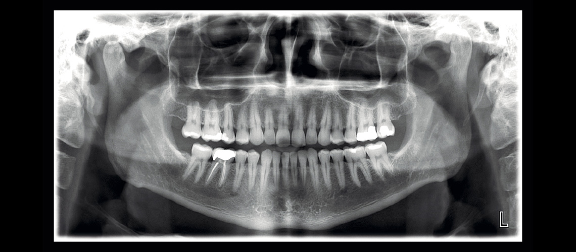
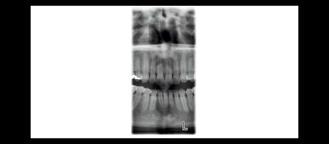
In addition to the Standard panoramic program, the following programs are offered: Interproximal panoramic program: generates an image, where interproximal teeth contacts are open. Primarily used for caries detection. Orthogonal panoramic program: produces an image with clearly visible alveolar crest for improved diagnostics. Ideal for periodontal imaging and implant planning.
Extraoral bitewings
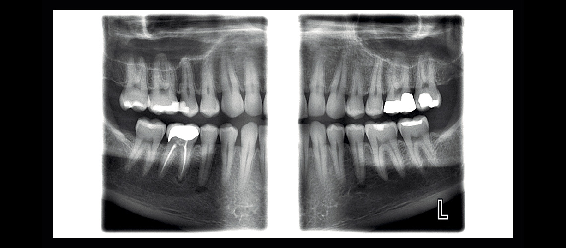
The Bitewing program uses improved interproximal angulation geometry. The result is a bitewing image pair with low patient dose and excellent diagnostic quality.
Sinus Imaging
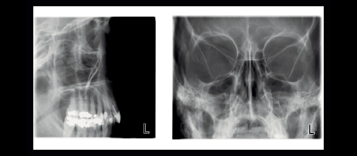
The Sinus programs provide a clear view of the maxillary sinuses.
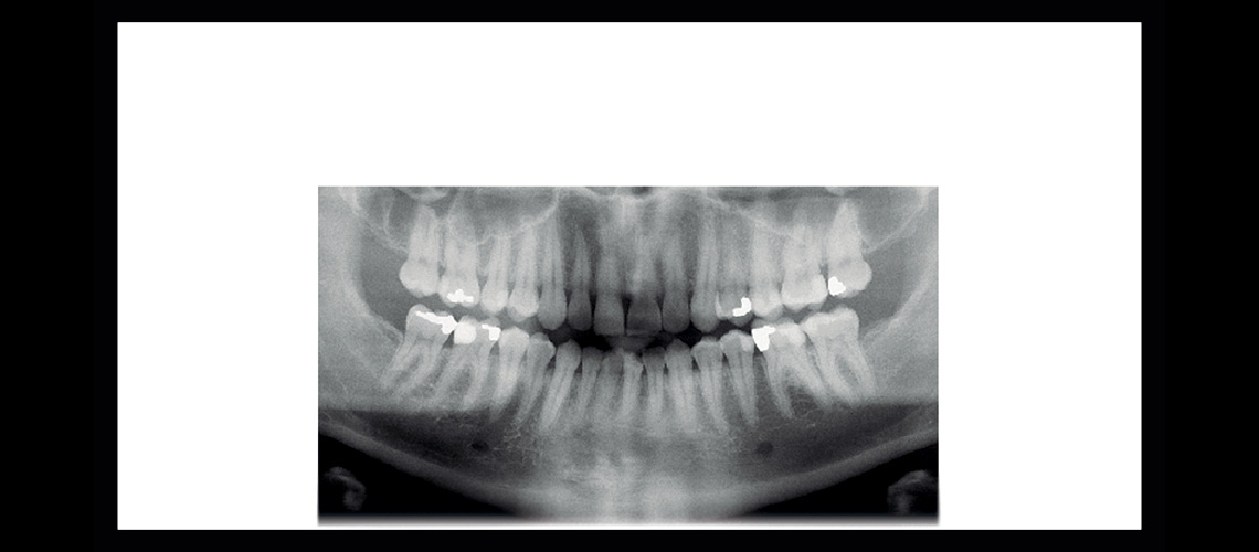
Horizontal and vertical segmenting for panoramic program With the Horizontal and vertical segmenting program, exposure can be strictly limited to the diagnostic region of interest. Patient dosage is reduced by up to 90% compared to full panoramic exposure.
TMJ Imaging
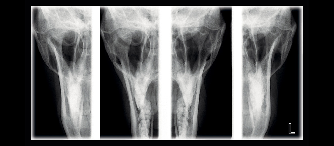
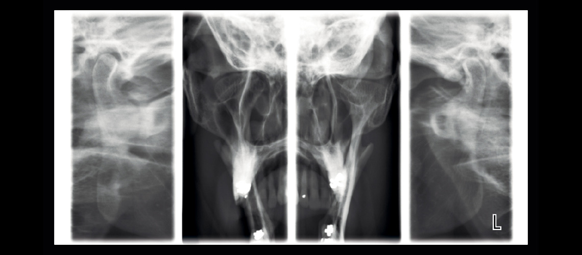
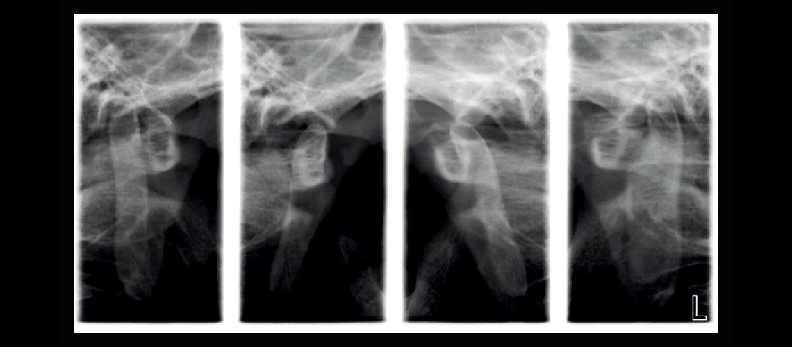
The TMJ imaging programs produce lateral or posteroanterior views of open or closed temporomandibular joints. The imaging angle and position can be adjusted to correspond to the anatomy of each individual patient. The Lateral-PA TMJ program captures lateral and PA views on the same radiograph. The multi-angle TMJ programs produce radiographs with images from three different angles, from either the lateral or PA view.
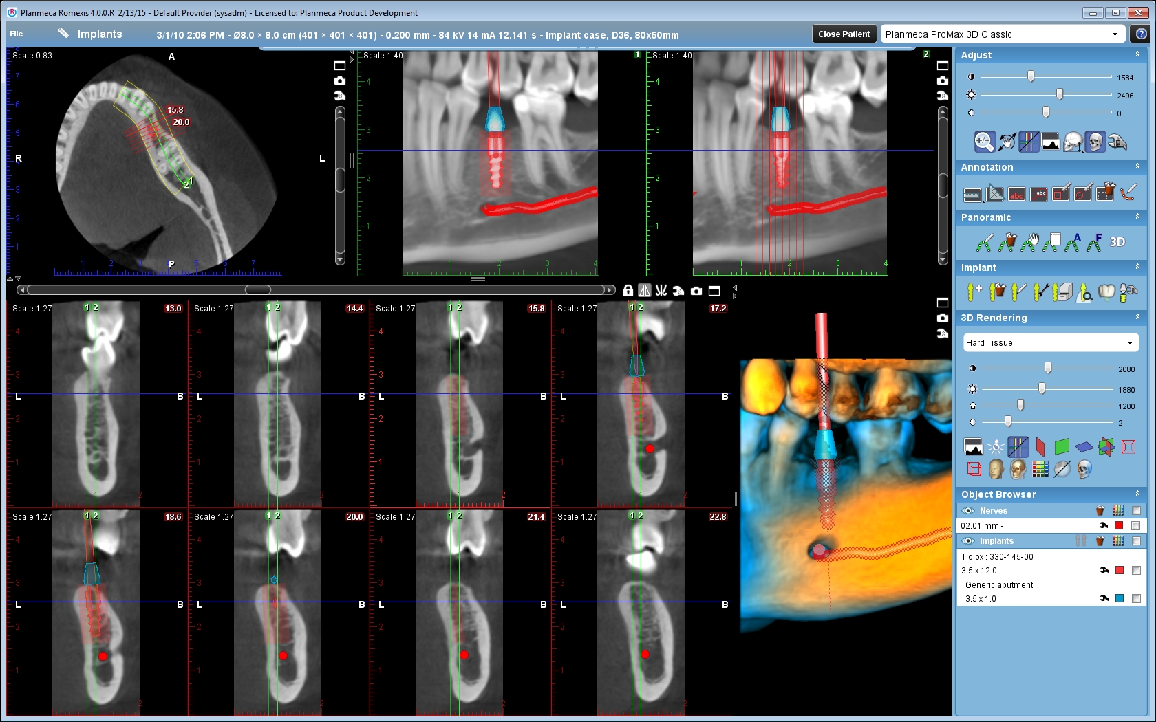
Easy upgrade from 2D to 3D
Planmeca ProMax® 2D is designed with upgradeability in mind. The unit’s modular structure allows easy conversion to different imaging modalities, while the software-driven SCARA is extremely flexible, allowing you to benefit from new imaging projections.
Whether you’re upgrading your 2D unit to 3D, or adding a cephalometric arm, Planmeca has the right solution for you. Individual options can be installed before delivery or added later, making Planmeca ProMax the most versatile all-in-one X-ray unit available.
